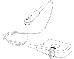
Ultrasound imaging is a non-invasive technique that uses sound waves to create images of the internal structures of the body. Ultrasound transducers are the devices that emit and receive the sound waves and convert them into electrical signals that are processed by a computer to form an image. Ultrasound transducers come in various shapes and sizes, depending on the purpose and the body part they are used for.
Some of the most common types of ultrasound transducers are:
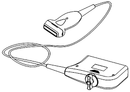
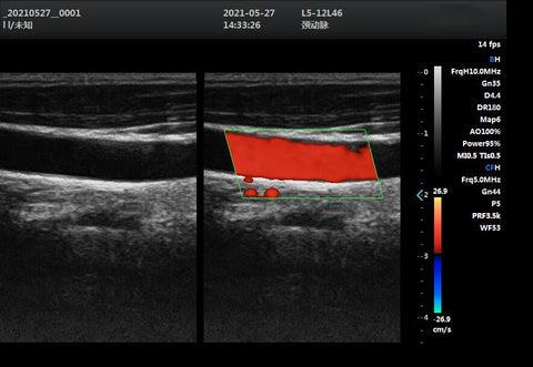
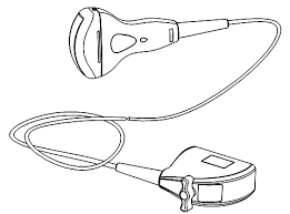
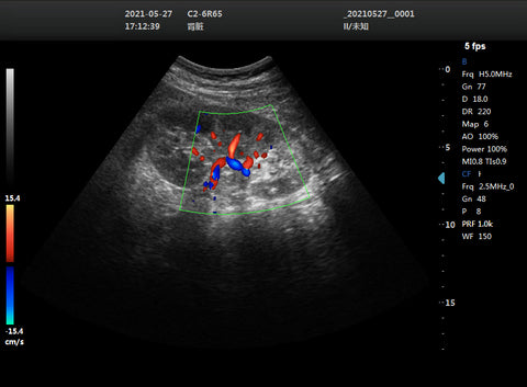
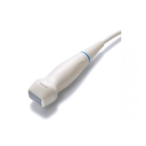
• Linear transducers: These have a linear arrangement of piezoelectric crystals that produce a rectangular beam of sound waves. They are used for imaging superficial structures, such as blood vessels, thyroid, breast, and tendons. They have a high frequency and a good resolution in the near field, but a poor resolution in the far field. • Convex transducers: These have a curved arrangement of piezoelectric crystals that produce a convex beam of sound waves. They are used for imaging deeper structures, such as abdominal organs, fetal development, and pelvic organs. They have a low frequency and a good resolution in the far field, but a poor resolution in the near field. • Phased array transducers: These have a small arrangement of piezoelectric crystals that are electronically controlled to produce a narrow beam of sound waves that can be steered in different directions. They are used for imaging hard-to-reach structures, such as the heart, brain, and lungs. They have a low frequency and a variable resolution depending on the beam angle and depth.
• Endocavity transducers: specialty transducers used to image structures from inside the body. This allows for better visualization of structures that are not easy to view with a surface transducer. The shape of the imaging surface provides a very wide field of view. Endocavity transducers are most frequently used for OB/GYN and urology applications.
• Volume transducers: specialty transducer that captures 3D/4D images and can be utilized in a variety of applications. They can be useful in helping clinicians to appreciate depth better and achieve a more realistic representation of anatomy in additional planes. Volume transducers are typically used for cardiac and OB/GYN imaging.
• Micro-convex transducers:generally employed for veterinarian, pediatrics and neonatal abdominal, vascular and cardiac applications. – Endocavitary: Endocavitary transducers are employed for women's health applications. This includes projecting urological, OB-GYN, endovaginal / transvaginal and fetal images.
There are also other types of ultrasound transducers that are designed for specific applications, such as transesophageal transducers, which are inserted into the esophagus, and pencil transducers, called CW Doppler probes, are utilized to measure blood flow and speed of sound in blood. This probe has a small footprint and uses low frequency (typically 2Mhz– 8Mhz).

 Ultrasound imaging is a non-invasive technique that uses sound waves to create images of the internal structures of the body. Ultrasound transducers are the devices that emit and receive the sound waves and convert them into electrical signals that are processed by a computer to form an image. Ultrasound transducers come in various shapes and sizes, depending on the purpose and the body part they are used for.